Analytical Assays For Endpoint Data Collection
Preclinical Histology & IHC Services
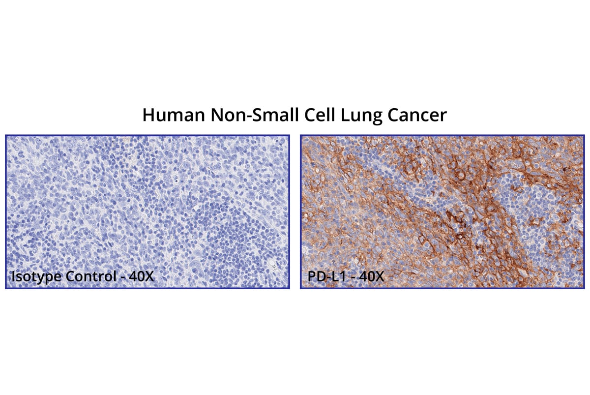
At Champions Oncology, we provide comprehensive histology services and immunohistochemistry (IHC) testing to support a wide range of research areas. Our advanced IHC markers enable the precise detection of key biomarkers, while our experienced team ensures each IHC procedure is conducted with the highest level of rigor and accuracy.
By integrating histology and IHC, we offer a holistic view of tissue architecture and protein expression, providing researchers with the detailed insights needed to drive preclinical discoveries. Our expertise in IHC pathology establishes a strong foundation for producing high-quality, reproducible data across diverse studies.
Capabilities
Custom Developed Histology & IHC Services
Expertise in preclinical sample analysis, including complex sample analysis needed for PDX models
GCLP-compliant clinical sample preparation and analysis
Over 200 custom developed protocols and fully optimized antibodies for research
Model Selection
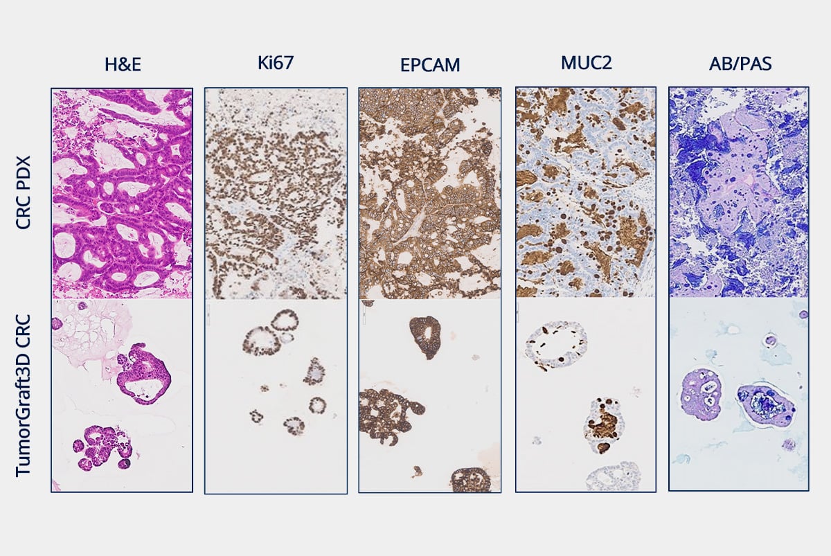
Confidently Select TumorGraft® Models through Specialized IHC
Clinical Samples
Evaluate Target Expression in Clinical Patient Samples With Our Histopathology Services
At a Glance
Champions Oncology's Histology and IHC Services
| Category | Description | Technology | Expertise |
|---|---|---|---|
| Histology Sample Preparation | Tissue collection, processing, FFPE generation | Automated staining and TMA | Board-certified pathologists |
| IHC | Antibody staining, target expression analysis | Optimized protocols, slide digitization | TumorGraft® models specialization |
| Tissue Imaging | Microscopic Imaging | Advanced imaging technologies | Preclinical and clinical research |
Histology and IHC services across the sample lifecycle, from preparation through imaging
Testimonials
"Their tissue analyses are high caliber and their dependability has significantly improved our capacity to gather insights and make informed decisions about our drug development initiatives."
VP of Drug Discovery, Pharmaceutical company
"Champions' Clinical IHC services are exceptional, providing detailed tissue staining and high-quality results essential for evaluating our therapeutic and its effects in a clinical setting."
Clinical Operations, Pharmaceutical Company
"Their tissue analyses are high caliber and their dependability has significantly improved our capacity to gather insights and make informed decisions about our drug development initiatives."
VP of Drug Discovery, Pharmaceutical company
FAQs
Frequently Asked Questions
Using preclinical histology services offers several advantages, including:
- Expertise: Access to specialized knowledge and skills in tumor tissue preparation and analysis.
- Advanced Technology: Utilization of state-of-the-art imaging and optimized staining techniques for precise results.
- Efficiency: Outsourcing histology tasks can accelerate project timelines by leveraging dedicated resources.
- Quality Control & Assurance: Ensures consistent and high-quality results that meet regulatory standards, supporting the credibility of preclinical findings.
Histology utilizes various imaging technologies to visualize and analyze tissue samples, including:
-
Light Microscopy: Commonly used for examining stained tissue sections at high resolution.
-
Fluorescence Microscopy: Allows for the visualization of specific molecules within tissues using fluorescent tags.
-
Confocal Microscopy: Provides detailed 3D images of tissues by scanning multiple planes within a sample.
-
Electron Microscopy: Offers ultra-high resolution images of tissue ultrastructure, revealing cellular details at the nanometer scale.
Resources
Explore our resources
- Leveraging IHC as a powerful clinical tool for IO therapiesBlog - February 16, 2023
- Histology & Immunohistochemistry (IHC)Platform Sheet
Related services
Explore related services