
<img alt="" src="https://secure.soil5hear.com/223550.png" style="display:none;">
Select Models
1715912769

Analytical Assays For Endpoint Data Collection

Preclinical In Vivo Imaging

Cutting-edge preclinical imaging services to assess the effectiveness of your cancer therapeutics, monitor disease progression, and study cancer metastasis

Overview

Champions' Unrivaled Preclinical In Vivo Imaging Services

Using preclinical in vivo imaging along with Champions' cutting-edge oncology research platforms, researchers can evaluate disease progression and metastasis formation in real time.

A growing portfolio of luciferase-tagged tumor models optimized for use in the preclinical in vivo imaging platform

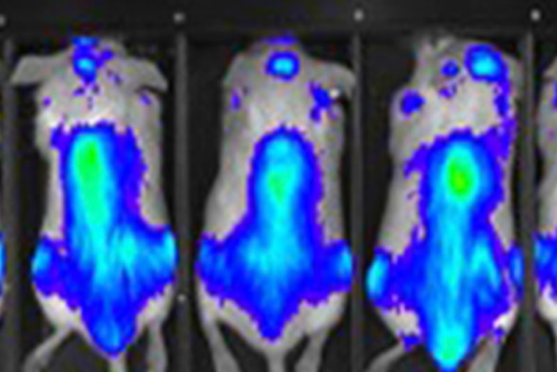
Leverage non-invasive bioluminescence imaging to measure tumor burden in real-time, including systemic disease models

Enables the visualization of metastasis and micro-metastasis, to assess the impact of therapeutic agents on metastatic processes

Preclinical in vivo imaging using IVIS provides real time analysis.
spect-ct

Radiopharmaceutical Imaging

In-House SPECT/CT Imaging

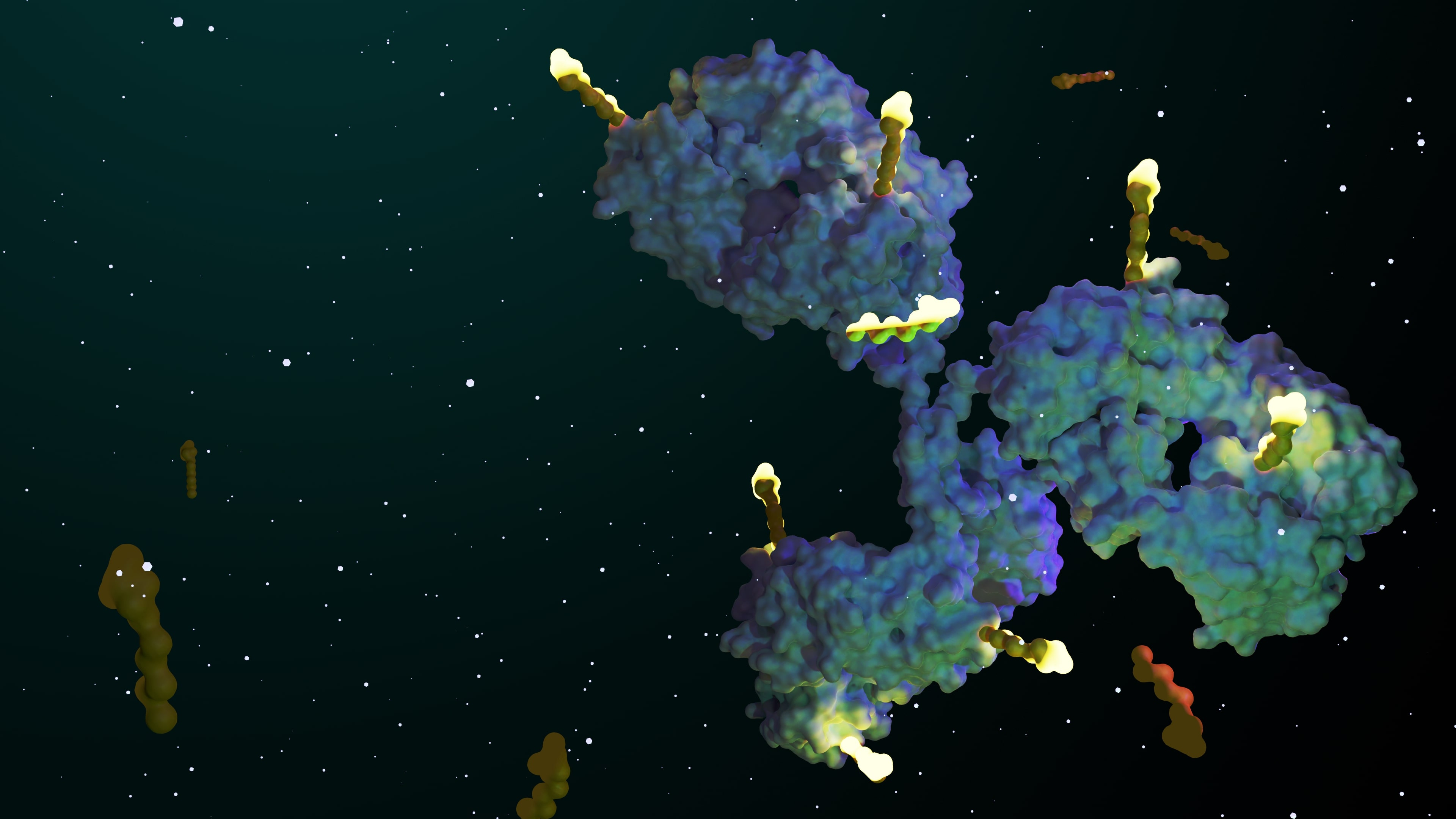
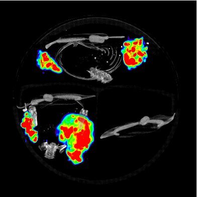
See exactly where your radiolabeled compound goes, and when. Champions' in-house Molecubes γ-X CUBE micro-SPECT/CT system delivers non-invasive, longitudinal imaging of tumor targeting and biodistribution for radiopharmaceutical studies, integrated directly with our CDX and PDX model catalog.

  • Quantitative outputs, including %ID/cc, standardized uptake value (SUV), and time-activity curve (TAC) analysis

  • Raw DICOMs and co-registered (fused) SPECT/CT images delivered alongside your study data

  • Runs in the same coordinated workflow as your radiolabeling, biodistribution, and efficacy studies, with one team from start to finish

For the full radiopharmaceutical development workflow, see our radiopharmaceutical services page. 

Imaging Platform

How Our Preclinical In Vivo Imaging Platform Works

Champions' Advantage

We pair preclinical in vivo imaging directly with our PDX and CDX model catalog, so imaging isn't a bolt-on service; it's built into the same study your models are already running in.

That means:

  • A growing, luciferase-tagged model portfolio ready for bioluminescence imaging out of the box
  • Non-invasive, longitudinal tracking that follows one animal over the full course of a study, reducing the number of animals needed
  • In-house SPECT/CT imaging for radiolabeled compound programs
Champions' Instrumentation

Preclinical in vivo imaging involves non-invasive techniques used to study biological processes in live animal models, such as mice and rats, before advancing to human clinical trials. This imaging approach is crucial for understanding disease mechanisms, assessing drug efficacy, and monitoring treatment responses in a controlled laboratory environment. 

Champions Oncology has integrated preclinical in vivo imaging into its advanced research platforms to enhance the study of oncology disease progression in systemic tumor models. By utilizing bioluminescence imaging (BLI), researchers can non-invasively track and measure tumor burden in real-time throughout an in-life study. Champions has optimized several luciferase-tagged tumor models within this platform, enabling precise detection and monitoring of tumor growth to evaluate therapeutic efficacy effectively. 


FAQ

Frequently Asked Questions

In vivo imaging is a non-invasive or minimally invasive technique that enables real-time visualization of biological processes within a living organism. It allows researchers to monitor disease progression, assess drug efficacy, and track cellular or molecular changes over time without the need for sacrificing the subject. In preclinical cancer research, preclinical in vivo imaging technology serves as a powerful tool for studying tumor biology and therapeutic responses in animal models like mice and rats. By providing quantitative, real-time insights into cancer progression and treatment effects, in vivo imaging enhances study precision while reducing animal usage through longitudinal assessments. 

Key Applications in Preclinical Cancer Studies

  • Tumor Growth & Metastasis Monitoring: Tracks tumor development over time in orthotopic (organ-specific) and systemic models and can help detect and quantify metastasis non-invasively.

  • Therapeutic Efficacy Assessment: Evaluates drug response, including tumor regression and relapse, and can measure treatment-induced changes in tumor size, metabolism, and molecular markers.

  • Pharmacokinetics & Drug Distribution: Analyzes how anti-cancer drugs distribute, metabolize, and clear within the body, and also helps optimize dosing and therapeutic windows.

  • Tumor Microenvironment & Immune Response: Visualizes immune cell infiltration and tumor-immune interactions and can study the impact of immunotherapies, such as checkpoint inhibitors and CAR-T cell therapies.

  • Angiogenesis and Hypoxia Studies: Assessing tumor blood vessel formation and oxygenation status can help researchers investigate anti-angiogenic therapies targeting tumor vasculature. 

Common In Vivo Imaging Modalities in Preclinical Cancer Research

  • Bioluminescence Imaging (BLI): Uses light-emitting luciferase-tagged cancer cells to monitor tumor growth and metastasis.

  • Fluorescence Imaging (FLI): Tracks labeled cancer cells, immune cells, or drug molecules.

  • Positron Emission Tomography (PET): Detects metabolic activity in tumors using radiolabeled tracers (e.g., FDG for glucose metabolism).

  • Magnetic Resonance Imaging (MRI): - Provides high-resolution images of tumor anatomy and vascularization.

  • Computed Tomography (CT): Generates detailed 3D tumor imaging, often combined with PET for anatomical context.

  • Ultrasound: Visualizes tumor growth and blood flow in real time.

  • SPECT/CT: Combines single-photon emission data with CT for precise anatomical localization of radiolabeled tracers; available in-house on our Molecubes γ-X CUBE system. 

Bioluminescence imaging (BLI) is a non-invasive optical imaging technique used to visualize and track biological processes in live animals. It relies on the emission of light from cells or tissues engineered to express luciferase, an enzyme that produces light when it reacts with its substrate (e.g., luciferin).

  • Propagation and in vitro characterization of luciferase-tagged cell lines

  • Systemic or orthotopic implantation into a live animal, such as mice or rat

  • Conduct pilot studies to study tumor growth kinetics

  • Monitor tumor growth by longitudinal imaging using IVIS

  • Design efficacy study testing various treatment modalities

  • Analysis using IVIS imaging software 

Yes, for radiolabeled compound programs. Our in-house Molecubes γ-X CUBE micro-SPECT/CT system is fully integrated with our radiochemistry and CDX/PDX model workflows, delivering non-invasive, longitudinal imaging of tumor targeting and biodistribution for radiopharmaceutical studies. It provides quantitative outputs including %ID/cc, standardized uptake value (SUV), and time-activity curve (TAC) analysis, plus raw DICOMs and co-registered SPECT/CT images. Visit our radiopharmaceutical development page for the full service details. 
1477214805

Get started

Get in touch

Fill out the form to learn about adding preclinical imaging as an endpoint for your in vivo studies.