<img alt="" src="https://secure.soil5hear.com/223550.png" style="display:none;">
Skip to content

Translational TMAs for Oncology Research

Accelerate biomarker discovery, validate targets, and benchmark therapeutic impact, all from a single, high-efficiency platform.

Champions Oncology's Patient Derived Xenograft (PDX) Tissue Microarrays (TMAs) deliver more than static samples. They provide a functional starting point for integrated oncology research. By offering hundreds of PDX tumor cores per slide, our TMAs eliminate bottlenecks in early-phase validation and help scientists quickly identify which targets matter. 

Tumor Microarray H&E staining slides.

Why Choose Champions Translational TMAs?

  • Patient-Derived and Clinically Annotated: Assembled from our TumorGraft™ and , our TMA cores reflect Western clinical treatment history and come annotated with genomic and histopathologic data. 
  • High-Content, Low-Variability Format: Each slide contains cores from a diverse patient cohort, reducing staining variability and enabling reliable IHC/IF analysis at scale. 
  • Ready for IHC, and Multiplexing: Built on FFPE format, our TMAs are compatible with standard histology workflows and multiplex biomarker readouts. 
  • Pipeline-Linked, Not Standalone: TMAs are directly traceable to Champions’ matched preclinical models, including PDX, and TumorGraft3D™ organoids, enabling immediate, functional follow-up. 

Translational Value Beyond the Slide  

Each array isn’t just a diagnostic snapshot, it’s a launchpad. When a marker or expression pattern is identified, Champions enables seamless progression into:

  • Functional testing in TumorGraft3D™ or co-culture assays
  • Drug efficacy and combination studies in PDX or ex vivo systems

Using a prostate cancer-specific TMA, a client validated AR expression patterns across resistant patient samples, then used matched ex vivo models to test a dual inhibitor strategy. The result: clear translational direction in weeks, not months. 

Her2 IHC staining of a breast cancer Tissue Microarrays (TMA)

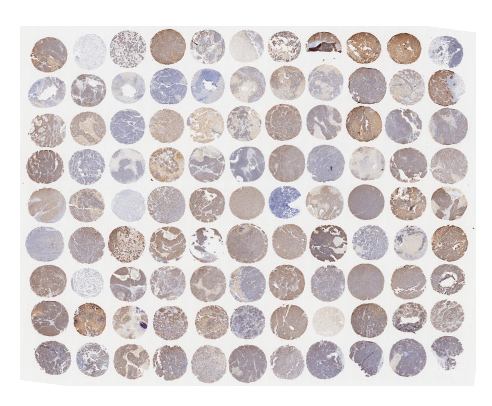
Her2 IHC staining of a breast cancer TMA

Key Applications

  • Antibody validation and optimization
  • Biomarker discovery and target expression mapping 
  • Indication stratification and tissue profiling 
  • Preclinical immuno-oncology screening and hypothesis testing 

Available Arrays

  • Broad coverage of solid and hematologic tumors 
  • Rare and treatment-resistant cancer types
  • Large inventory available off-the-shelf 
  • Preclinical immuno-oncology screening and hypothesis testing 

Request Our TMA Catalog 

or contact our scientific team to explore available tissue types and matched preclinical models.

Frequently Asked Questions
What is the tumor microarray technique?
The tumor microarray (TMA) technique involves extracting small, cylindrical tissue cores from  tumor blocks and embedding them in a single recipient paraffin block. This allows simultaneous histological analysis of hundreds of samples under identical experimental conditions.
What is a tissue microarray used for?
Tissue microarrays are primarily used for high-throughput biomarker screening, target validation, and comparative tissue profiling. They help researchers analyze large cohorts of tumor samples efficiently and cost-effectively.
What can microarrays tell us about cancer?
TMAs can reveal patterns of protein expression, target prevalence, and biomarker distribution across diverse tumor types and patient samples. This enables insight into tumor heterogeneity, resistance markers, and therapeutic relevance.
What are the disadvantages of tissue microarray?
While TMAs offer scalability and consistency, they may underrepresent intratumoral heterogeneity due to the small core size. Additionally, sample dropout or damage during processing can occasionally limit full slide utility.
How are Champions’ TMAs different from other providers?
Champions' Translational TMAs are directly linked to functional tumor models (PDX, ex vivo, TumorGraft3D™), enabling follow-up validation beyond the slide. Our arrays are also clinically annotated and reflect Western treatment profiles, making them more relevant for translational research.
What types of cancers are included in Champions’ TMAs?
We offer arrays spanning common and rare solid tumors, as well as hematologic malignancies. Indications include breast, prostate, lung, colorectal, melanoma, ovarian, DLBCL, and more.
Can I perform immunohistochemistry (IHC) or multiplex staining on Champions’ TMAs?
Yes. All of our TMAs are FFPE-based and compatible with standard IHC, , and multiplex staining protocols.
Can TMAs be used for antibody validation?
Absolutely. TMAs are an ideal tool for antibody specificity and sensitivity validation across diverse tumor backgrounds before transitioning to in vivo or functional studies.
How do I transition from a TMA hit to a functional study?
Each TMA core in our platform is traceable to matched preclinical models. Once a target is identified, our scientific team can guide you into follow-up studies using PDX, ex vivo, or 3D multiclonal assays.
How can I request a custom TMA or view your catalog?
You can contact our team to request a full catalog or discuss a custom array design based on your research needs and preferred indications.